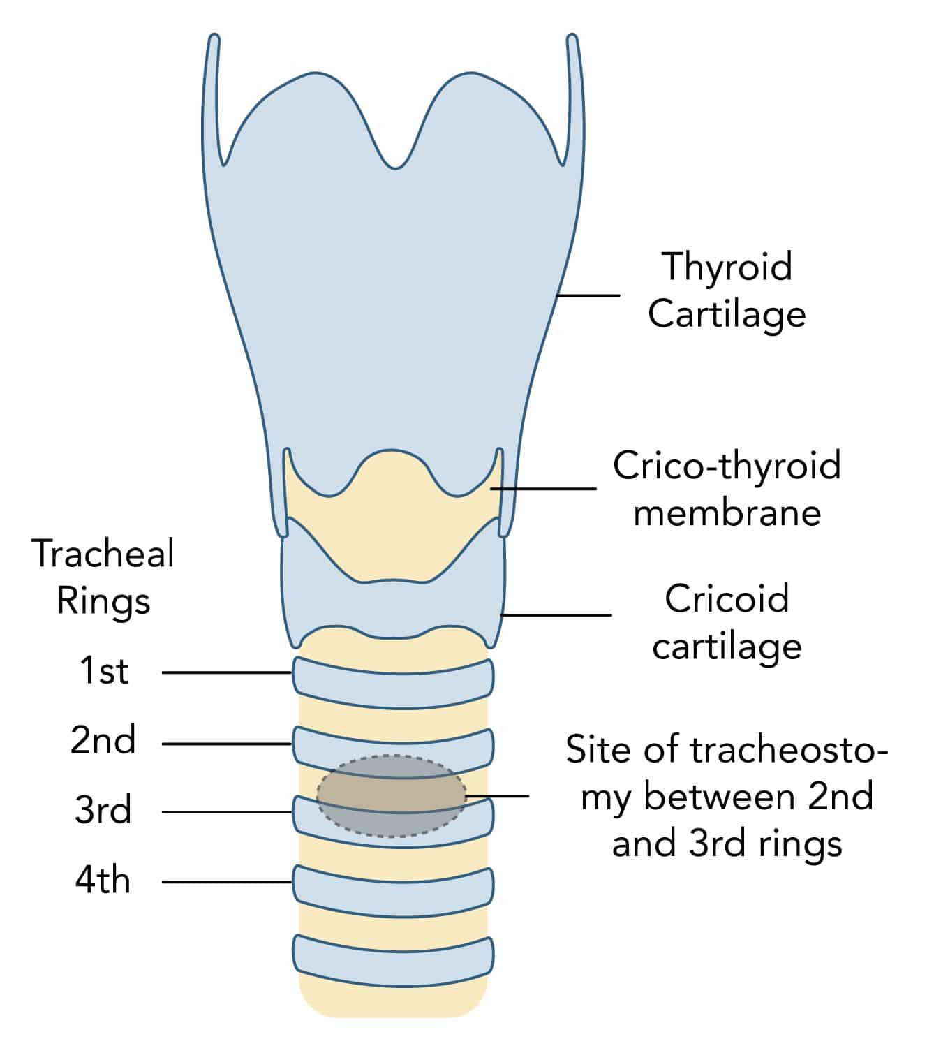
What is the preferred site of insertion for percutaneous tracheostomy and why?
- Preferred site of tracheostomy is between the 2nd and 3rd tracheal rings in the midline
- Tracheostomy above this site:
- Can lead to damage to the cricoid cartilage and first tracheal ring
- Results in increased risk of subglottic stenosis which is difficult to treat
- Tracheostomy below this site:
- Can lead to damage to the thyroid and great vessels at the root of the neck
- Results in increased risk of significant bleeding
What is the structure and course of the trachea?
- Tube of cartilage with a membranous lining continuous with the larynx
- Composed of 16–20 C-shaped cartilaginous rings
- Trachealis muscle completes the posterior wall
- Around 10-12 cm in length
- Extends down from cricoid at C6
- Terminates at carina at T5-
- Moves anteriorly to posteriorly from the cricoid distally
- Enters the chest behind the sternal notch
Which vessels are at risk of damage during tracheostomy?
- Anterior jugular veins:
- Run vertically close to the midline
- Thyroid ima artery :
- Ima is ‘lowest’ in Latin
- Anatomical variant in 3–10% of the population
- More common British Asian populations
- Arises mainly from the brachiocephalic trunk and ascends along the front of the trachea
- Inferior thyroid veins
- Other vessels more lateral: internal jugular vein, carotid artery, external jugular vein
What is the relationship of the thyroid to the trachea?
- Lobes lie laterally to the trachea and extend to the 6th tracheal rings
- Isthmus crosses the midline in the region of the 2-4th tracheal rings
- Pyramidal lobe, an anatomical variant, may extend superiorly from the isthmus to the cricoid


