Basics of TOE
Indications, Contraindications and Complications
What are the contraindications to transoesophageal echocardiography?
Absolute contraindications
- Patient refusal
- Active upper GI bleeding
- Esophagectomy
- Recent oesophageal surgery
- Upper GI perforation
- High risk oesophageal pathology:
- Stricture
- Tumor
- Diverticulum
Relative Contraindication
- Peptic ulcer disease
- Lower risk oesophageal pathology:
- Varices
- Barrett’s oesophagus
- Oesophagitis
- Dysphagia
- Cervical spine restriction
- GI surgery
- Recent upper GI bleeding
- Laceration
- Radiation to neck or chest
- Coagulopathy
- Symptomatic hiatal hernia
- Thoracoabdominal aneurysm
What are the complications of transoesophageal echocardiography?
Complication
Incidence
- Lip Injury
- Hoarseness
- Dysphagia
- Major bleeding
- Arrhythmia
- Death
- Laryngospasm
- Bronchospasm
- Oesophageal perforation
- Heart failure
- Tracheal intubation
- Pharyngeal injury
- Odynophagia
- Dental injury
Scanning Protocol & Views
Sequence & Summary
What are the sequence of views used in a basic TOE exam?
- Previously 20 views recommended for comprehensive examination
- Realistic to expect focus on at least 11 most relevant views:
1. Mid-Oesophageal Four-Chamber View
2. Mid-Oesophageal Two-Chamber View
3. Mid-Oesophageal Long-Axis View
4. Mid-Oesophageal Ascending Aortic Long-Axis View
5. Mid-Oesophageal Ascending Aortic Short-Axis View
6. Mid-Oesophageal AV Short-Axis View
7. Mid-Oesophageal RV Inflow-Outflow View
8. Mid-Oesophageal Bicaval View
9. Transgastric Midpapillary Short-Axis View
10. Descending Aortic Short-Axis View
11. Descending Aortic Long-Axis View
What are the features of the 11 key views used in a basic TOE exam?
30-35cm
- Advance slowly
- Turn clockwise or counterclockwise to centre on the mitral valve.
- Right atrium
- Interatrial septum (IAS)
- Left atrium
- Left ventricle (inferoseptal wall on left, anterolateral wall on right)
- Right ventricle
- Interventricular septum
- Mitral valve (A3A2 and P2P1 scallops)
- Tricuspid valve (posterior and septal leaflets)
- Chamber size (LA, RA, LV, RV)
- Global and regional (inferoseptal and anterolateral walls) LV systolic function
- Global and regional (lateral wall) RV systolic function
- Interatrial septum for presence of shunts
- MV function (morphology and colour doppler)
- TV function (morphology and colour doppler)
Midoesophageal Four-Chamber View
How should the probe be positioned for the ME four-chamber view
- Advance slowly to a depth of approximately 30-35 cm until it is immediately posterior to the left atrium
- Rotate the multiplane angle to approx 0-20°:
- Aortic valve (AV) or LV outflow tract (LVOT) should no longer be displayed
- Maximise the tricuspid annular dimension (often involves rotating to 15°
- Turn the probe to the left (counterclockwise rotation) or to the right (clockwise rotation of the probe) is performed to centre the mitral valve MV) and left ventricle in the sector display.
How should the image be optimised for the ME four-chamber view?
- Adjust the depth to ensure viewing of the left ventricular (LV) apex.
- Slight retroflexion may be required to align the MV and LV apex and prevent foreshortening of the ventricle.
- Turning the probe to the left (counterclockwise) allows imaging of primarily the left heart structures.
- Turning the probe to the right (clockwise) allows imaging of primarily right heart structures.
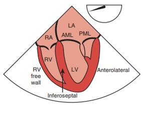
What image is seen in the ME 4-chamber view?
What image is seen in the ME 4-chamber view?
• Interatrial septum (IAS)
• Left atrium
• Left ventricle (Inferoseptal wall on left, anterolateral wall on right)
• Right ventricle
• Interventricular septum
• Mitral valve (A3A2 and P2P1 scallops)
• Tricuspid valv (posterior and septal leaflets)
• Coronary sinus (after slight probe advancement, imaged in the long-axis)
• Left upper pulmonary vein (after turning the probe to the left (anticlockwise) and withdrawing slightly)
What are the advantages of using ….. in the assessment of ….?
What are the limitations of using ….. in the assessment of ….?

