Time: 0 second
SOE 492: Pancreatitis
Question No. 2
Q: What is acute pancreatitis and how is it defined?
Answer No. 2
- A common presentation and a surgical emergency
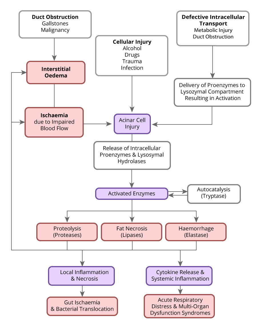
- Results from an inflammatory process in the pancreas leading to a multisystem disease characterized by a systemic inflammatory response and multi-organ dysfunction syndrome
Pancreatitis is typically established by the presence of two of the following criteria:
1. Abdominal pain consistent with the disease
2. Serum amylase and/or lipase greater than three times the upper limit of normal
3. Characteristic findings from abdominal imaging
Question No. 3
Q: How can severity of acute pancreatitis be classified?
Answer No. 3
Pancreatitis can be classified by severity according to the Revised ATLANTA criteria 2012
Mild
- No organ failure
- No local complications
Moderate
- Transient organ failure (<}48 hours)
Severe
- Local complications +/-
- Persistent organ failure
*Local complications include:
Acute peripancreatic fluid collection
Pancreatic pseudocyst
Acute necrotic collection
Pleural effusion
Question No. 4
Q: What is the mortality rate of severe acute pancreatitis?
Answer No. 4
- Still carries high mortality though overall deaths remain stable despite increased incidence:
- Reflects recent advances in management
- Mortality rate of up to 30% reported
Question No. 5
Q: What are the causes of pancreatitis?
Answer No. 5
Obstructive / Mechanical
- Gall stones (50%)
- Malignancy:
- Pancreatic ductal carcinoma
- Ampullary carcinoma
- Islet cell tumour
- Sarcoma
- Lymphoma
- ERCP (5% of procedures)
- Trauma
- Penetrating duodenal ulcer
- Congenital abnormalities
- Cystic fibrosis
Parenchymal
- Alcohol (20%)
- Autoimmune / vasculitic disease
- Scorpion stings
Systemic
- Hypoxemia / ischaemia
- Drugs
- Hypothermia
- Hypercalcemia
- Hypertriglyceridemia (>20mmol/L)
- Viruses (Mumps, HIV, CMV, EBV)
- Other infections
- Venovenous - requires an external pump
MEMORY TIP
A commonly used mnemonic is: I GET SMASHED
- Idiopathic
- Gall stones (50%)
- Ethanol (20%)
- Trauma
- Steroids
- Mumps and other viruses (EBV, CMV, HIV)
- Autoimmune diseases (SLE, polyarteritis nodosa, pregnancy)
- Scorpion stings
- Hypercalcemia, hyperlipidemia, hypothermia, hypotension (ischemia)
- ERCP, emboli
- Drugs
Question No. 7
Q: What is the role of Imaging in pancreatitis?
Answer No. 7
- An ultrasound of the RUQ should be performed in all cases to assess for biliary stones and obstruction
- After negative routine work-up for biliary aetiology, endoscopic ultrasonography (EUS) is recommended as the first step to assess for occult microlithiasis, neoplasms and chronic pancreatitis
- If EUS is negative MRCP is advised as a second step to identify rare morphologic abnormalities
Question No. 8
Q: Which scores can be used to assess the severity of pancreatitis?
Answer No. 8
- A number of systems exist to identify severity and prognosticate pancreatitis
- Considered advantageous over clinical judgement alone
- Useful in determining optimum location of care
- Limitations exist with many of the scoring systems:
- Cumbersome to complete
- Require 48 hours to gather variables for some scores
- Lack accuracy in early stages
- Limited clinical value
Classification Systems
Atlanta Criteria
- Divides pancreatitis in to two pathophysiological types:
- Interstitial oedematous pancreatitis
- Necrotising pancreatitis
- Classifies severity as mild, moderate and severe
- Determined by presence of local features and organ failure
Prognostic Scoring Systems
Disease Specific
Clinical
- Ransoms:
- Originally designed for gallstone-induced pancreatitis
- Uses age, nine laboratory parameters plus fluid requirements to calculate a score over 48 hours
- A score of >3 at 48 hours indicates the presence of severe pancreatitis
- Glasgow-Imrie:
- Requires 48 hours to complete
- Uses age and seven laboratory parameters to predict severe pancreatitis
- BISAP
Radiological
- Balthazar CT grade
Non-Specific
- APACHE II
- A score of >8 at 24 hours defines severe acute pancreatitis
Question No. 9
Q: What are the complications associated with acute severe pancreatitis?
Answer No. 9
Local (Pancreatic)
- Interstitial Oedematous pancreatitis:
- Peripancreatic collection
- Pseudocyst (pancreatic fluid surrounded by a wall of fibrous or granulation tissue)
- Abscess (circumscribed collection of pus)
- Necrotising pancreatitis:
- Sterile parenchymal necrosis
- Infected parenchymal necrosis
- Pancreatic insufficiency (and Type 3c Diabetes)
Regional
- Ascites
- Portal vein / splenic thrombosis
- Intraperitoneal bleeding
- Retroperitoneal bleeding
- Bowel obstruction
- Enteric fistulas
- Intrabdominal hypertension
Systemic
- Sepsis
- Multiorgan failure
- Respiratory:
- Respiratory failure
- ARDS
- Effusions
- DIC
- Renal failure
- GI Bleeding
Question No. 10
Q: When should a patient with acute pancreatitis be managed within critical care?
Answer No. 10
- UK guidelines state that all patients with acute severe pancreatitis should be managed on the high dependency unit (HDU):
- Can be difficult in practice - ensure all referred to the critical care outreach team for regular review and escalation of care if deterioration occurs
- Particular features which suggest critical care admission may be beneficial include:
- Age 70 years or older
- Body mass index over 30 kg/m2
- Hypotension not responsive to fluid resuscitation
- >30% necrosis of the pancreas
- Pleural effusions
- Three or more of Ranson's criteria
- CRP > 150 mg/L at 48 hours
(Adapted from World Association Guidelines)
Question No. 11
Q: How would you manage the patient with severe acute pancreatitis?
Answer No. 11
Key Principles
- Aggressive fluid resuscitation and pain management
- Early ERCP if indicated
- Early enteral feeding (preferably via NG route)
- Avoid early surgical intervention for necrotic pancreatitis
- Vigilant supportive care to avoid complications
Initial Resuscitation & Supportive Care
- ABCDE approach treating abnormalities as found
- Manage airway and breathing:
- If intubation may be required, aspiration a major risk
- Multifactorial causes for respiratory failure (ARDS, diaphragmatic splinting (pain, intra-abdominal oedema or fluid collections) or pleural effusions)
- Optimise haemodynamics:
- Early and aggressive fluid resuscitation
- May require vasopressor support if severe systemic inflammatory response
- Maintain UO >0.5 ml/Kg
- Manage electrolyte abnormalities:
- Vigilance over hypocalcaemia and arrhythmias
- Ensure optimal analgesia:
- PCA usually required
- Some support use of thoracic epidural
- Aim to prevent further atelectasis
- Correct coagulopathy in the setting of VTE
- Optimise nutrition:
- Early enteral feeding (within 72 hours):
- Nasogastric - effective in 80%
- Nasojejunal second line
- No advantages for early TPN - only after 7 days if enteral fails
- Early enteral feeding (within 72 hours):
- Vigilance to good supportive care - often long stays and prone to complications
- Strict glycaemic control
- DVT prophylaxis (balance against risk of intrabdominal haemorrhage)
- Stress ulcer prophylaxis
- VAP bundles
- Aseptic precautions
Specific Management
- Management of biliary obstruction
- Early ERCP indicated to remove gallstone (within 72 hours)
- Coagulation should be corrected
- Surgical management of gallstones during same hospital admission or within 2 weeks
- Management of infected collection / necrosis
- Prophylactic antibiotics not routinely recommended
- If clinical sepsis ensure blood cultures taken and treat as per surviving sepsis
- If >30% pancreatic necrosis, should undergo FNA to obtain material for culture 7–14 days after the onset of the pancreatitis
- If infected abscess confirmed post-needle aspiration prescribed according to local guidelines
- If infected will require definitive intervention (Ideally delay until 4 weeks):
- Radiological drainage first line - successful in 50%
- Endoscopic drainage
- Surgical drainage (delay until clear demarcation)
- If evidence of retroperitoneal gas on CT:
- Broad spectrum antibiotics
- Surgical drainage or debridement
- Delayed surgery (>2 weeks):
- Associated with increased survival
- Allows demarcation of necrotic and preserved tissue
Referral & Deposition
- All patients with sever acute pancreatitis should be manages on HDU
- Refer all with persisting organ failure or requiring intervention to regional centre
Question No. 12
Q: What nutritional therapy should patients with severe acute pancreatitis receive?
Answer No. 12
- Early EN is now standard of care in patients with acute pancreatitis
- Recent research suggests improved outcomes compared with previous strategies of pancreatic rest with TPN
- Guidance recommends commencing within 72 hours if intolerant of oral intake
- Enteral nutrition in acute pancreatitis can be administered via either the nasojejunal or nasogastric route:
- Many recommend early NJ feeding and supported by ESPEN guidance
- Two trials have also found no difference in outcomes in patients fed gastrically versus jejunally
- Nasogastric usually successful in 80% of patients
- Nasojejunal feeding should be used if intra-abdominal pressures are >15mmHg
- Parenteral nutrition can be administered in acute pancreatitis as second-line therapy if nasojejunal tube feeding is not tolerated (pain, ileus, nausea)
(NICE & ESPEN Guidelines)
Question No. 13
Q: Which patients should receive antibiotics in severe acute pancreatitis?
Answer No. 13
- Antibiotics should be used in any case of pancreatitis complicated by infected pancreatic necrosis but should not be given routinely for fever, especially early in the presentation:
- Carbapenem usually the best class due to penetration in to pancreatic tissue
- Fungal infection should be considered in severe infected pancreatic necrosis
- Image-guided FNA should be used to gain material for culture when:
- Necrosis and features of sepsis
- 30% necrosis and persistent symptoms at 7-14 days
- Antibiotic prophylaxis in severe pancreatitis is controversial:
- Trials performed in this area show great heterogeneity with a variety of antibiotics used for different durations
- Routine use of against infection is not currently recommended
- If used may increase the risk of fungal superinfection
Question No. 14
Q: What are the indications for surgical and radiological interventions in severe acute pancreatitis?
Answer No. 14
- Relieving biliary obstruction (e.g. ERCP)
- Removing infected intra- and extra-pancreatic necrosis (Necrosectomy)
- Pancreatic duct disruption
- Management of symptomatic masses due to pseudocysts or sterile necrosis:
- Gastric outlet or intestinal obstruction
- Persistent pain

