Time: 0 second
Question No. 2
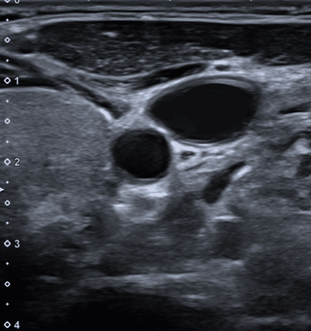
Q: Which side of the patients neck is this image of? (1 marks)

Answer No. 2
- This is the right side 1 of the neck
- The thyroid gland can be identified medially
- The internal jugular vein lies laterally to the carotid artery

1
Question No. 4
Q: Other than the internal jugular vein what are the contents of the carotid sheath? (3 marks)
Answer No. 4
- Vagus nerve 1
- Carotid artery 1
- Deep cervical lymph nodes 1

3
Question No. 5
Q: What are the classical landmarks for insertion of an IJV CVC using the landmark technique? (3 marks)
Answer No. 5
Technique
Classical (Central)
- Insertion point is apex of Sedillot's triangle 1
- Sternal head of sternocleidomastoid 1
- Clavicular head of sternocleidomastoid 1
- Superior border of medial 3rd of the clavicle 1
- Needle angled at a 45° to the skin 1
- Aiming toward the ipsilateral nipple 1

3
Question No. 10
Q: Where is the appropriate position for an internal jugular CVC tip on CXR? (1 marks)
Answer No. 10
- The most commonly used x-ray landmark is the level of the carina 1:
- Origin of the Superior vena cava 1-2cm above
- Pericardial reflection lies 1-2cm below
- Tip should lie within the boundaries
- Cavo-atrial junction usually lies two vertebral bodies below the carina:
- Above this may be an acceptable position for L sided lines to ensure they lie parallel or haemodialysis catheters to optimise flow

1